Scan #5 | 16 Months Since Dx

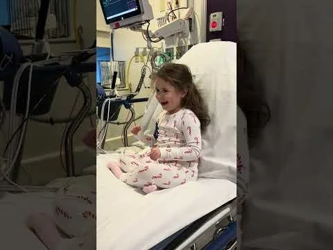
Another complicated scan.
Some quick good news, first: the hemorrhagic lesion identified on our last scan has not changed at all (that’s good!) and there are no signs of tumors in Juliette’s spine.
Now the concerning update: the hyperintensity we’ve been tracking has changed, and we’re not sure if it’s the tumor growing again or scar tissue. The radiology report notes the lesion now demonstrates a nodular associated enhancement with a small focus of probable decreased diffusivity along its periphery. Findings are worrisome for residual/recurrent tumor rather than postsurgical changes given the temporal change in size and signal characteristics.
“Nodular associated enhancement” - this means that the hyperintensity absorbed more contrast than it has previously, indicating increased bloodflow to that area which can signal tumor or scar tissue.
“Decreased diffusivity” - this means that there’s an increase in cell density, again typically signaling tumor or scar tissue.
Unfortunately there is no test to distinguish tumor from scar tissue. The only way to know with certainty is for a neurosurgeon to resect the tissue and have it analyzed.
Next Steps:
Juliette will have a lumbar puncture performed in the next couple weeks. The CSF (cerebrospinal fluid) obtained via the lumbar puncture will be analyzed for ctDNA. This test is sophisticated and fairly new. It could detect cancer before the MRI.
Juliette will receive another MRI scan in February to track the size and behavior of the hyperintensity.
Juliette’s most recent scans will be reviewed by the neurosurgical team at Boston Children’s Hospital in order to establish a recommendation, which could include brain surgery.